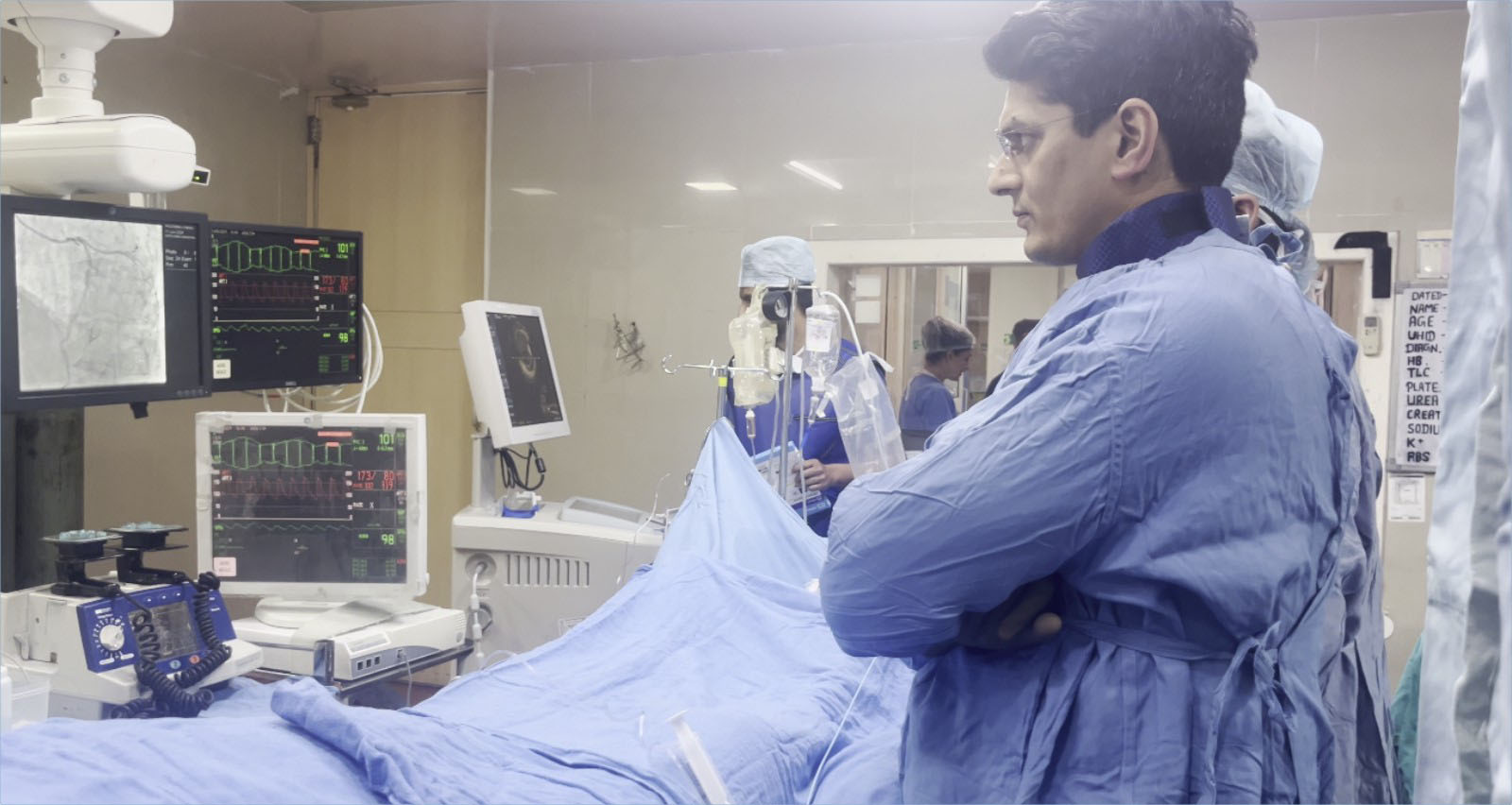
Dr Rakesh Sharma

what is coronary Angiography

Angiography is a medical imaging modality that depicts the interior of blood vessels and organs. The procedure involves the administration of a contrast agent into the vessels, thereby making them more visible during imaging scans. Thus, here is a more elaborative overview:

Types of Angiography

  • Coronary Angiography
    Purpose: This is aimed at viewing the coronary arteries, vessels that supply blood to the myocardium.
    How it’s done: A thin, flexible tube called a catheter is inserted through an artery in the groin or wrist and threaded to the coronary arteries. Contrast dye is delivered, allowing visualization of blood flow to reveal any blockages or narrowing.
    Indication: Mainly taken for the diagnosis of CAD and intervention, like angioplasty or the placement of a stent, and also for assessing the condition of the heart.

  • Peripheral Angiography
    Purpose: To visualize the blood vessels outside the heart, usually in one’s legs or arms.
    Procedure: Like coronary angiography, except this focuses on peripheral arteries. It assists a doctor in diagnosing peripheral artery disease (PAD) and assessing the extent of blockage in the blood vessels.

  • Carotid Angiography
    This allows providing images of the carotid arteries in the neck that feed blood to the brain.
    How it’s done: A catheter is usually inserted into the femoral artery, located in the groin, then threaded to the carotid arteries. Contrast dye is then injected to detect any blockage or narrowing that may cause strokes.

  • Pulmonary Angiography
    To visualize the blood vessels in the lungs.
    A catheter is inserted into the pulmonary arteries, followed by the administration of contrast dye to outline the blood flow within the vessel. It is commonly utilized for diagnosing any vascular condition in the lungs but most specifically for pulmonary embolism.

  • Renal Angiography
    It is used for viewing, through the use of contrast dye, the renal vessels.
    Procedure: Administration of contrast dye into the renal arteries views its functionality of the kidneys and locates any tumor formation or area of stenosis.
    How it is done: Patients are usually asked to withhold food for a few hours prior to the examination. A sedative or local anesthesia is frequently used.

  • Contrast Administration:
    The catheter is progressed through the blood vessel, and a contrast agent is administered intra-vascularly. The dye then delineates the blood vessel on X-ray or other imaging modalities.
    Imaging studies such as X-rays, CT scans, or MRI may be done to take pictures of the blood vessels. Images obtained are then studied for abnormalities.

  • Post-procedure
    The patient is observed for immediate complications. He or she can be asked to lie flat for a couple of hours if the catheter insertion is via the groin.
    Diagnosis: Blockages, aneurysms, or other abnormalities of blood vessels.
    Treatment planning: To assist the doctor in choosing the best method for surgeries or interventions.
    Follow-Up: Monitoring the effectiveness of treatments or interventions.

  • Risks
    Allergic Reaction: Rarely, patients can be allergic to the contrast dye.
    Bleeding: There is an outside chance of bleeding or infection at the site of catheter insertion.
    Kidney Damage: On rare occasions, the contrast dye can affect kidney function.
    Angiography is a beneficial modality in modern medicine, extending valuable assistance in many vascular pathologies regarding their diagnosis and management.

We TAKE CARE of You from our HEART.

Book Appointment with Dr Rakesh